What is Age-Related Macular Degeneration (AMD)?
A common eye condition and leading cause of vision loss amongst people over 50, age-related macular degeneration, or AMD, is deterioration or damage of the macula, a small area near the center of the retina. The macula is comprised of light-sensing cells that provide your central vision. The retina then turns the light into electrical signals and sends them through the optic nerve to the brain where they are translated into the images we see. The center of your field of vision may appear blurry or distorted when the macula is damaged.
Risk Factors
AMD is most likely to occur after the age of 60 but other major factors leading to its onset can include:
Smoking: According to the American Academy of Ophthalmology (AAO), smoking doubles the risk of AMD.
Race: Age-related macular degeneration is most common among Caucasians.
Family history: Patients with a family history of AMD are at a greater risk of developing it.
How is AMD detected?
The early stages of AMD typically begin without symptoms. A comprehensive dilated eye exam will detect signs of AMD as it provides a better view of the back of your eye. An ophthalmologist may also perform a fluorescein angiogram. In this test, a fluorescent dye is injected into your arm. Pictures are taken of the dye as it passes through the blood vessels in your eye which allow your doctor to see leaking blood vessels, which are the hallmark for severe AMD.
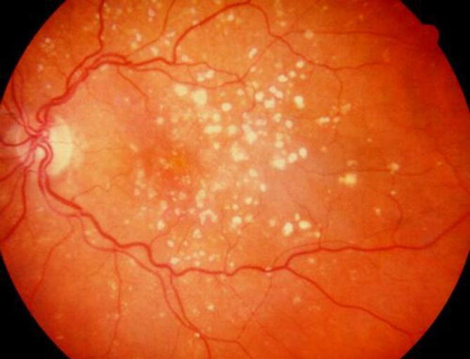
Another sign of AMD is the appearance of drusen which are yellow deposits beneath the retina. The presence of medium to large drusen is a strong indicator of AMD as it develops in stages.
The Stages of AMD
Early AMD is diagnosed by the presence of small drusen which are approximately the width of a human hair. Vision loss typically does not occur at this time.
Intermediate AMD in patients is usually determined by the appearance of medium to large drusen and/or pigment changes in the retina. At this stage, patients still may not experience symptoms despite the possibility of slight vision loss.
Late AMD is divided into two categories:
Geographic atrophy (Dry AMD): a gradual breakdown of the light-sensitive cells in the macula that transmit visual information to the brain.
Neovascular AMD (Wet AMD): is the growth of abnormal blood vessels underneath the retina which can leak fluid and blood, damaging the macula.
It is very possible to have both types of late AMD in the same eye as either condition can appear first. Not all patients with early AMD will develop late AMD. However, people who have early AMD in one eye and no signs of AMD in the other eye; 5% of those will develop late AMD after 10 years. Having late AMD in one eye increases the risk of it developing in the other eye.
How is AMD treated?
Early AMD: Since many patients show no signs of vision loss at this stage, there are no treatments. That being said, it is imperative that patients under a comprehensive dilated exam once a year to determine if AMD is advancing. The preventative measures that patients can take for early AMD are frequent exercise, avoiding smoking and eating nutritious foods such as leafy vegetables and fish.
Intermediate and Late AMD: According to the National Eye Institute, nutritional supplements (AREDS and AREDS2) along with vitamins and minerals can slow the progression of AMD.


Contact Information
Office Hours
- Monday 9:00 AM - 6:00 PM
- Tuesday 8:00 AM - 6:00 PM
- Wednesday 8:00 AM - 7:00 PM
- Thursday 8:30 AM - 6:00 PM
- Friday 8:00 AM - 5:00 PM
- Saturday 8:00 AM - 3:00 PM
- Sunday Closed
Optical Store Hours
- Monday 9:00 AM - 6:00 PM
- Tuesday 9:00 AM - 6:00 PM
- Wednesday 9:00 AM - 6:00 PM
- Thursday 9:00 AM - 6:00 PM
- Friday 9:00 AM - 5:00 PM
- Saturday 8:00 AM - 3:00 PM
- Sunday Closed
© 2026 The Center for Eye Care and Optical. All rights Reserved. Accessibility Statement - Privacy Policy - Sitemap
Powered by: