


articles
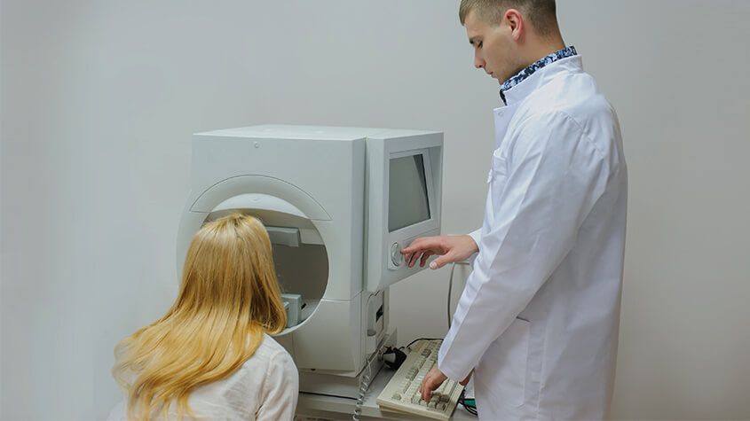
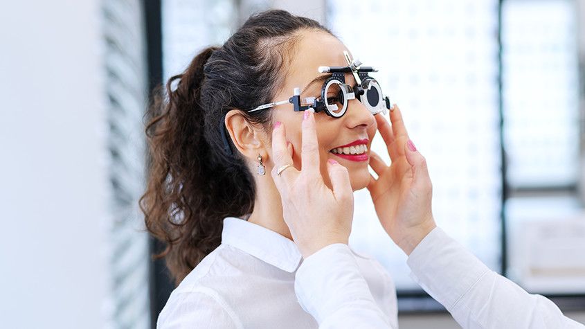
Visual field testing is an important part of most standard comprehensive eye exams. Also sometimes known as perimetry testing, Visual field testing is a method to measure the entire scope of vision of an individual, including their peripheral/side vision.
The importance of visual field testing
Visual field testing is one of the most effective diagnostic treatments in the detection of glaucoma. This is because when patients are affected by glaucoma, it is usually the peripheral vision that is affected by their condition first. However, it can also be used to detect central or peripheral retinal diseases, eyelid conditions such as drooping, optic nerve damage and conditions that affect the visual pathways from the optic nerve to the area of the brain where this information is processed into vision.
Visual field testing is also an important part of monitoring for people who are considered to be at risk for vision loss from disease and other problems, including those who have been diagnosed with the following:
Multiple sclerosis
Hyperthyroidism
Pituitary gland disorders
Central nervous system problems (such as a tumor that may be pressing on the brain)
Stroke
Diabetes
High blood pressure
What to expect from visual field testing
There are a variety of methods that can be used to perform visual field testing, including:
Static automated perimetry. This is where a machine is used to quantify how well the patient is able to detect flashing lights of varying size and brightness in different areas of their visual field, while they concentrate on a central point. The patient responds by pushing a button when they see the light.
Kinetic perimetry. This involves points of light that are fixed in size and intensity and are presented along the patient’s peripheral vision, before being gradually moved inwards to determine their field of vision.
Visual field testing is non-invasive, painless and doesn’t require patients to have their eyes dilated. The results, which are usually presented in a series of charts, are digital and sent directly to your eye doctor for interpretation. Depending on the outcome of your results, you may be recommended for further diagnostic testing which could include blood tests. If you have been diagnosed with glaucoma, you will probably be recommended to have several visual field tests each year, which will help your eye doctor to monitor the progression of your condition and recommend treatments to slow it.
If you would like more information about visual field testing, or if you have concerns about your peripheral vision, please don’t hesitate to schedule an appointment with our experienced and knowledgeable eyecare team today.
Millions of people suffer from eye and vision-related problems. For some, the solution is as simple as wearing prescription eyeglasses or contact lenses. However, others face challenges that aren’t as easy to rectify. That’s where Lumenis comes into play.
As a world leader in minimally invasive clinical solutions for both ophthalmology and aesthetic markets, it’s making a significant difference. Not only does Lumenis develop advanced energy-based technologies, but it also commercializes them.
The company continues to find innovative solutions. However, it’s already provided the fields of ophthalmology and aesthetic with remarkable technologies. OptiLight is just one example.
Eye and Vision-Related Solution
The company invented the first and only technology to treat individuals for dry eye disease caused by MGD. Not only is it approved by the Food and Drug Administration (FDA) but also patented. Optimal Pulse Technology (OPT) makes it easier to successfully manage this particular eye disease. Overall, OptiLight is safe, precise, comfortable, and effective.
Managing Symptoms
According to recent statistics, roughly 22% of the U.S. population suffers from dry eye disease along with MGD. That combination leads to an array of uncomfortable symptoms.
Dry eye occurs when a person’s eyes don’t produce adequate tears to keep them lubricated or when tears don’t work the way they should. MGD is a condition that affects the small glands in the eyelid responsible for making the oil layer for tears.
Having to deal with one of these problems is bad enough. However, living with both can prove debilitating for some people. Thanks to OptiLight, people with the two conditions get much-needed relief. Here are the ways this advanced technology helps:
Reduces inflammatory mediators, which, thereby, prevents inflammation
Improves tear breakup time that, in response, decreases osmolarity
Alleviates abnormal blood vessels that commonly cause inflammation
Restores meibomian glands so they function properly
Decreases demodex mites that cause infection and the accumulation of bacteria on the eyelids
What Does OptiLight Consist Of?
As part of this revolutionary system, ophthalmologists and optometrists can select different devices based on their needs.
Patented OPT Handpiece
Not two people have identical faces. Even among twins, facial contours differ, even if slightly. To ensure excellent results, this handpiece allows ophthalmologists and optometrists to customize treatments to cover every curve.
Ergonomic IPL Handpiece
To treat a broader area, Lumenis recommends this option. Patented with SapphireCool technology, it breaks the cycle of inflammation associated with dry eye disease caused by MGD.
Opti-Tip
For maximum energy control, the Opti-Tip focuses light energy on more delicate areas. As with the other Lumenis devices, it’s safe and effective. At the same time, the Opti-Tip is 100% hygienic.
Key Benefits of OptiLight
OptiLight utilizes embedded settings that adhere to strict protocols, making it safe and effective tool.
Additionally, they use Optimal Pulse Technology (OPT), which transforms light-based therapy. As a result, ophthalmologists and optometrists can treat the eyes with absolute precision and control. That’s because while this technology provides optimal energy, it never has spike inconsistencies.
Doctors can treat a patient in just 15 minutes. Specifically for dry eye disease with MGD, it provides an improvement in just four sessions.
The Bottom Line
Lumenis dedicated years to develop OptiLight. Now, eye doctors can incorporate a system into their practice to provide better patient care.
Ocular aesthetics, which focuses on improving the appearance of the eyes and the surrounding area, has gained significant popularity in recent years. Ocular aesthetics aims to rejuvenate the eyes, making them appear more youthful, vibrant, and refreshed. This can be achieved through advanced technology treatments that are designed to address specific concerns and enhance the overall appearance of the eyes.
Common Concerns in Ocular Aesthetics
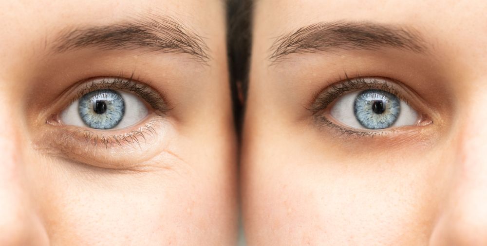
There are several common concerns that individuals seek to address through ocular aesthetics. These concerns may be a result of natural aging, genetics, or lifestyle factors. One of the most common concerns is the appearance of wrinkles and fine lines around the eyes, commonly known as crow's feet. These lines can make you look older and tired.
Another common concern is the presence of dark circles and puffiness under the eyes. This can give the impression of fatigue and detract from the overall attractiveness of the eyes.
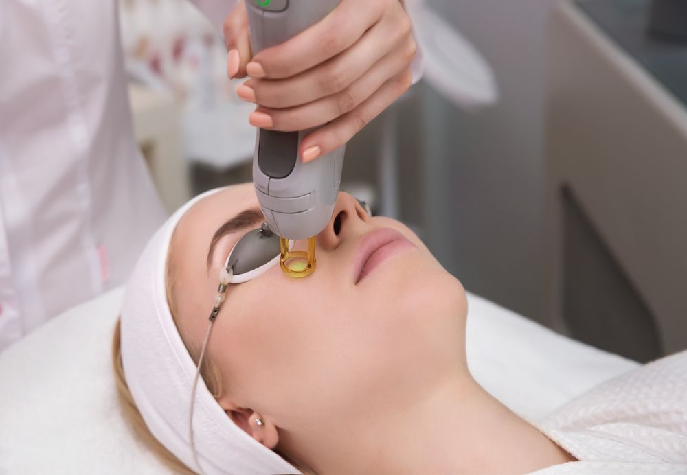
IPL and How it Works
Intense Pulsed Light (IPL) has emerged as a popular and effective treatment option in ocular aesthetics. IPL works by emitting high-intensity light pulses that target specific chromophores in the skin, such as melanin and blood vessels. This targeted approach allows for precise treatment of various concerns around the eyes.
IPL treatments can effectively address pigmentation irregularities, such as age spots and sun damage, which often contribute to an aged appearance. By targeting these concerns, IPL can help restore a more even skin tone and improve the overall aesthetics of the eyes.
IPL treatments are non-invasive and typically require minimal downtime. The number of sessions needed may vary depending on the specific concerns being addressed. By consulting your optometrist, you can determine the best treatment plan to achieve your desired ocular aesthetics.
Radiofrequency (RF) for Ocular Aesthetics
Radiofrequency (RF) treatments have gained significant popularity in ocular aesthetics due to their effectiveness in addressing various concerns around the eyes. RF works by delivering controlled radiofrequency waves into the deeper layers of the skin, stimulating collagen production and tightening the tissues.
One of the primary concerns that RF treatments can address is drooping eyelids. As we age, the skin around the eyes can lose elasticity, causing the eyelids to sag. RF treatments can effectively tighten the skin, resulting in a more youthful and lifted appearance. This can help restore the natural contour and symmetry of the eyes, enhancing their overall aesthetics.
RF treatments can also target under-eye bags, which can make the eyes appear tired and aged. By stimulating collagen production, RF can improve the overall tone and texture of the skin, reducing the appearance of under-eye bags and giving the eyes a more refreshed and vibrant look.
Exploring Low-Level Light Therapy (LLLT)
Low-Level Light Therapy (LLLT) has gained recognition in ocular aesthetics for its ability to promote cellular activity and stimulate collagen production. LLLT utilizes specific wavelengths of light that penetrate the skin, triggering a series of biological responses that can improve the health and appearance of the eyes.
LLLT treatments can effectively address concerns such as fine lines, wrinkles, and dark circles around the eyes. By stimulating collagen production, LLLT can help reduce the appearance of these concerns, resulting in a more youthful and vibrant look.
In addition to its anti-aging benefits, LLLT can also improve the overall health of the eyes. By promoting cellular activity, LLLT can enhance circulation, reduce inflammation, and support the natural healing process. This can be particularly beneficial for individuals with conditions such as dry eyes or eye fatigue.
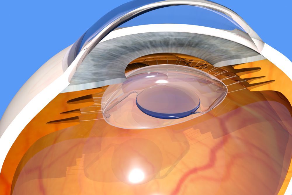
Clear Lens Extraction (CLE) is a surgical procedure that involves the removal of the natural lens of the eye and replacing it with an artificial lens implant, also known as an intraocular lens (IOL). CLE is primarily performed to correct refractive errors, such as nearsightedness, farsightedness, and astigmatism.
Understanding CLE and How It Works
Clear Lens Extraction is similar to cataract surgery, where the natural lens is removed and replaced with an IOL. However, in CLE, the lens is clear and not clouded as in the case of cataracts. The procedure begins with the administration of local anesthesia to numb the eye and ensure a painless experience for the patient.
Once the eye is numb, a small incision is made on the cornea, which is the clear front surface of the eye. Through this incision, your eye surgeon accesses the lens and carefully removes it. The artificial IOL is then inserted into the empty lens capsule. The IOL is specifically chosen to correct the patient's refractive error, providing them with improved vision.
After the IOL is implanted, the incision is closed with tiny sutures or self-sealing techniques. Following the surgery, patients are usually prescribed antibiotic and anti-inflammatory eye drops to prevent infection and reduce inflammation.
Factors to Consider Before Undergoing Clear Lens Extraction
Before deciding to undergo Clear Lens Extraction, there are several factors that need to be considered. Firstly, it is crucial to have a thorough eye examination to determine if you are a suitable candidate for the procedure. Your optometrist will evaluate your overall eye health and discuss your expectations and goals for vision correction.
Age is another important factor to consider. CLE is typically recommended for individuals over the age of 40 who have developed presbyopia, a condition that affects near vision. It is also important to have stable vision, as any changes in prescription can affect the accuracy of the IOL power calculation.
Additionally, it is essential to understand the potential risks and complications associated with the procedure. While CLE is generally safe, there is a small risk of infection, bleeding, and retinal detachment. Your optometrist will discuss these risks with you and address any concerns you may have.
Thanks to the advancement of lens technology, glasses lenses are no longer a single, one size fits all solution. There are a variety of different lens types that can be used in glasses, giving patients greater flexibility and control over their vision than ever before.
Single Vision Lenses
Also known as monovision lenses, these lenses are designed to correct the wearer’s vision at just one distance, and have a single prescription covering the entire surface of the lens. They are most often recommended for people who are either nearsighted (myopia) or farsighted (hyperopia) and who need glasses for a specific activity, such as driving or reading.
Progressive Lenses
Progressive lenses are multifocal lenses that can correct a patient’s vision at different working distances, ranging from far distance to reading distance. However, rather than designating different areas on the lenses for different distances with visible lines separating them, progressive lenses have a gradual change so that the wearer can smoothly transition from one lens power to another.
Bifocal and Trifocal Lenses
As you may have guessed from the name, bifocal and trifocal lenses have either two or three lens powers depending on which type you choose. Bifocal lenses support distance vision in the top half of the lens, and near vision in the lower half. Trifocal lenses support distance vision in the top third of the lens, intermediate vision in the middle segment and near vision in the bottom third. Whichever variety you choose, you will see visible lines separating each segment.
Bifocal and trifocal lenses are recommended for patients who are near or farsighted, and those who develop presbyopia, which is the natural hardening of the eye lens, that occurs as we get older. Presbyopia makes it harder for the lens of the eye to adapt to focus at different distances.
Multifocal Lenses
Multifocal lenses are the alternative name given to bifocal, trifocal and progressive lenses.
Computer Lenses
Computer lenses are prescription lenses that are specifically designed to be worn when doing computer work. This is because they place the optimum lens power for viewing your computer screen exactly where you need it – which is closer than intermediate vision, but further away than reading material is usually held. Wearing computer lenses can significantly reduce the negative effects caused by the high visual demands of computer work, including blurred vision, redness, dry eyes, double vision and dizziness.
Transition Lenses
Also known as photochromic lenses, transition lenses are a special type of lens that darken when in the sunlight and lighten when in softer light or the dark. This versatility gives the wearer the convenience of being able to move between different environments without needing to change their glasses. This makes them extremely cost effective and prevent the wearer from needing to take multiple pairs of glasses out with them. Transition lenses also filter out many of the harmful UV rays that are emitted from the sun, helping to keep eyes healthy too. They are ideal for people who spend a lot of time going between inside and outside, or who work outside in varying weather conditions.
Blue Light Lenses
Blue light lenses are specially crafted lenses that contain filters that block out much of the artificial blue light that is produced by digital devices like computers, smartphones and tablets. Natural blue light is actually good for balancing our sleep-wake cycle, boosting our mood and enhancing our cognitive abilities so that we can function better day to day. However, too much blue light, especially from artificial sources, can have the opposite effect. Many people who fail to use blue light lenses can go on to develop digital eye strain, which produces symptoms like eye fatigue, dry eyes, blurred vision, headaches and more. Blue light lenses are recommended for anyone who spends a lot of time working on a digital device.
Polarized Lenses
Polarized lenses are used to reduce eyestrain and improve the quality of vision in patients on especially sunny days, making them ideal for anyone who spends a lot of time outdoors. They can do this because they have a special filter that blocks some of the light from passing through the lens. Vertical light is allowed to pass through, while horizontal light, such as that which bounces off of water and can be blinding, is blocked. Polarized lenses are most often used in sunglasses since they are worn outdoors, and the wearer also needs to protect their eyes from UV damage.
Still have questions about which lens is right for you? Contact us to schedule an eye exam or an appointment to evaluate your individual needs.
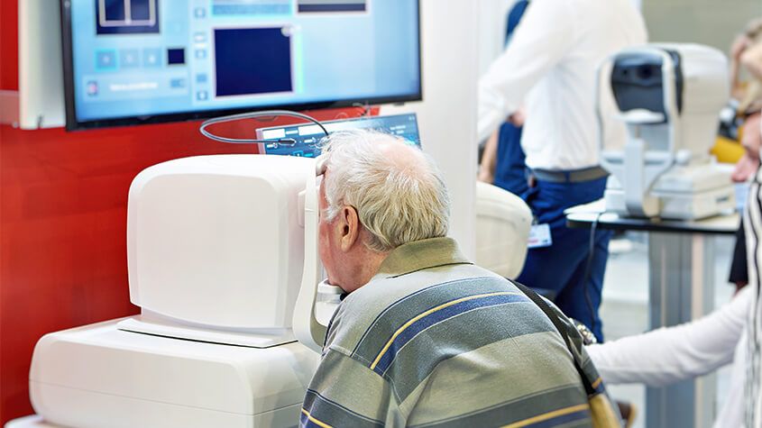
As technology continues to advance, so does the field of the optometric industry. The development of innovative tools and techniques has allowed for more accurate and comprehensive examinations. One such technology is Optos, a revolutionary system that utilizes ultra-widefield retinal imaging technology to provide optometrists with a detailed view of the entire retina.
How Does Optos Work?
Optos technology is based on the principle of ultra-widefield retinal imaging, which allows for a wider and more detailed view of the retina compared to traditional imaging techniques. The Optos system consists of a specialized camera that captures high-resolution images of the retina using scanning laser ophthalmoscopy (SLO) and optical coherence tomography (OCT) technologies. SLO provides a wide-field view of the retina, while OCT allows for cross-sectional imaging, providing valuable insights into the various layers of the retina.
The Optos camera is designed to capture images through a process called optomap, which captures up to 200 degrees of the retina in a single image. This wide-field view provides optometrists with a comprehensive picture of the retina, enabling them to detect abnormalities that may not be visible with traditional imaging techniques. The optomap image can be instantly viewed and analyzed by your eye doctor, allowing for a more efficient and accurate diagnosis.
Common Eye Conditions Detected by Optos
Optos technology has the capability to detect a wide range of eye conditions, including but not limited to, diabetic retinopathy, macular degeneration, glaucoma, and retinal tears or detachments. Diabetic retinopathy is a condition that affects individuals with diabetes, causing damage to the blood vessels in the retina. Optos can capture detailed images of the retina, enabling optometrists to detect any signs of diabetic retinopathy and initiate appropriate treatment.
Macular degeneration is another common eye condition that can be detected using Optos. This condition affects the macula, the central part of the retina responsible for sharp, central vision. Optos allows for a comprehensive view of the macula, identifying any changes or abnormalities that may indicate the presence of macular degeneration.
Glaucoma, a condition characterized by damage to the optic nerve, can also be detected using Optos. The wide-field view provided by Optos allows for a thorough examination of the optic nerve and the surrounding structures, facilitating early detection and intervention.
Finally, Optos technology is particularly effective in detecting retinal tears or detachments. These conditions can lead to sudden vision loss and require immediate medical attention. Optos allows for a comprehensive view of the retina, identifying any signs of retinal tears or detachments and initiate prompt treatment.

Dry eye is a common ocular condition that occurs when the eyes do not produce enough tears or when the tears evaporate too quickly. This can lead to discomfort, redness, blurred vision, and even damage to the surface of the eyes. Understanding the causes and symptoms of dry eye is crucial provide early detection and effective treatment.
The Importance of Dry Eye Advanced Diagnostic Testing
Early detection of dry eye is crucial for preventing further progression of the condition and improving patient outcomes. Without proper diagnosis and treatment, dry eye can cause significant discomfort and affect daily activities. Additionally, chronic dry eye can lead to corneal ulcers, infections, and even vision loss. By accurately identifying and addressing dry eye in its early stages, optometrists can provide timely interventions and prevent complications.
TearLab
One of the advanced diagnostic tools available for dry eye is TearLab. TearLab is a non-invasive test that measures the osmolarity of tears, which is an indicator of tear film stability. This test provides valuable information about the quality and quantity of tears, allowing healthcare professionals to accurately diagnose and monitor dry eye. By analyzing the osmolarity of tears, TearLab helps identify the severity of dry eye and guides treatment decisions. It is a quick and painless procedure that can be performed in a clinical setting.
InflammaDry
Inflammation plays a significant role in dry eye, and identifying the presence of inflammation is crucial for effective treatment. InflammaDry is a diagnostic tool that detects elevated levels of matrix metalloproteinase 9 (MMP-9), an inflammatory marker, in tears. By measuring MMP-9, InflammaDry helps optometrists differentiate between inflammatory and non-inflammatory dry eye. This information is essential for tailoring treatment plans and determining the most appropriate therapies for each patient.

Dry eye is a common condition that affects millions of individuals worldwide. It occurs when the eyes do not produce enough tears or the tears evaporate too quickly. This can lead to discomfort, irritation, and even vision problems. TearCare offers a breakthrough treatment option for long-lasting relief from dry eye.
What is TearCare?
TearCare is a revolutionary treatment option that offers long-lasting relief for dry eye sufferers. This innovative technology utilizes wearable eyelid warming devices to precisely deliver heat to the meibomian glands, effectively unclogging them and promoting healthier oil production. The treatment is performed in-office by a trained eye care professional and typically takes less than 15 minutes.
TearCare targets the root cause of the problem - meibomian gland dysfunction. By restoring the proper function of these glands, TearCare helps to stabilize the tear film, improve tear production, and alleviate the symptoms of dry eye. This breakthrough solution offers a safe and effective alternative to medications and artificial tear drops, providing long-lasting relief for those suffering from dry eye.
How TearCare Works to Treat Dry Eye
TearCare works by gently applying heat to the eyelids, stimulating the meibomian glands and promoting the release of healthy oils. The wearable eyelid warming devices are designed to maintain a consistent temperature, ensuring optimal results. The heat helps to liquefy any hardened oils or debris clogging the glands, allowing them to function properly again. This treatment can also help reduce inflammation and improve overall eyelid hygiene.

Wearing contact lenses gives patients the flexibility and freedom to live life to the fullest, without some of the difficulties presented by wearing glasses. Many people who choose contact lenses do so because they don’t like the way that glasses look or feel, or because wearing glasses compromises their ability to perform certain tasks or activities, such as sports or jobs that require the use of safety goggles.
There are lots of different contact lenses to choose from, with two of the most popular being daily disposables and toric lenses.
Disposable Lenses
As their name suggests, these daily contact lenses are disposable. This means that they can and should be discarded at the end of each day rather than re-worn. Disposable lenses do tend to be a little more expensive than some repeat-wear varieties, but the benefits usually outweigh the cost.
Some of the advantages of choosing daily disposable contact lenses include:
You don’t have to clean them, which saves patients a great deal of time and hassle. It also helps save money in terms of the ongoing cost of cleaning solution.
Disposable lenses are also great for people with eye allergies. This is because with ordinary lenses, there’s an opportunity for deposits and microorganisms to build up. With daily disposables, allergens have less chance to attach themselves to the lenses and cause irritation and other allergy symptoms.
You don’t need to schedule regular replacements either, which makes wearing contact lenses easier on your schedule.
Disposable contact lenses are particularly good for people who have busy lives and are likely to cut corners when it comes to caring for their eyes or contacts since there is no cleaning or maintenance required.
Daily disposable contact lenses are available in a wide range of prescriptions, including those for patients with nearsightedness and farsightedness. Your eye doctor will be able to advise you if you are a candidate for disposable contact lenses.
Toric Lenses
Toric contact lenses are recommended for patients who have a refractive eye problem called astigmatism. Patients with astigmatism have corneal abnormalities that cause the refraction of the eye to be different between the vertical and horizontal planes, causing blurred vision and difficulty seeing fine details. Toric contact lenses are shaped in a particular way that creates the different focusing powers needed in each part of the lens to correct your vision. For this reason, it’s essential that Toric lenses are placed into the eyes in the correct position.
Fortunately, manufacturers design Toric lenses with features that help them to stay in place, including:
Thin/thick zones
Creating areas of the lens that are thicker or heavier which helps secure it in position
An area where the bottom of the lens is slightly cut off
To keep them stable, Toric lenses are a little firmer than conventional soft lenses. This means that some patients can find them a little less comfortable, but the superior vision they obtain outweighs this. Your eye doctor will be able to advise you if you are a good candidate for Toric contact lenses and which variety would best suit you.
To find out more about daily contact lenses, speak to our friendly and knowledgeable team.
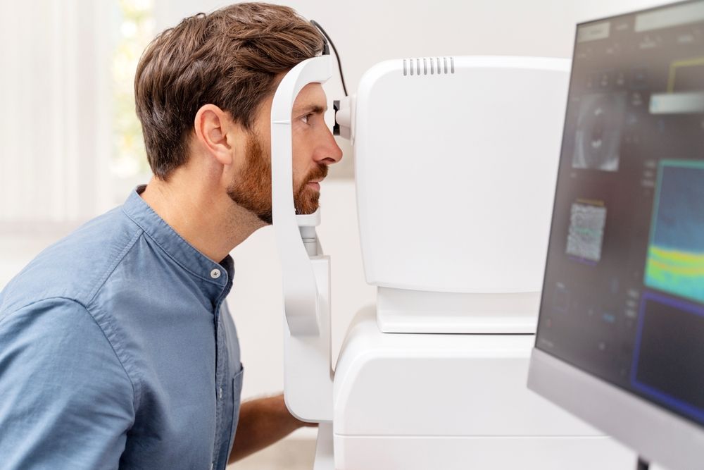
Optical Coherence Tomography is a non-invasive imaging test that may be performed as a standard part of your regular, comprehensive exams, or you may be able to request this test as an addition to your usual exam.
Optical Coherence Tomography uses light waves to take cross-section images of your retina, which is the area of light-sensitive cells at the back of your eye that is responsible for receiving light and transmitting it into messages that are sent up to the brain. The technology behind OCT enables your eye doctor to see each of the different layers that make up the retina. By being able to see these and measure them, they can obtain a much clearer picture of the overall health and condition of your eyes.
Why are Optical Coherence Tomography scans important?
When you choose to have an OCT scan at fairly regular intervals, such as during your normal comprehensive eye exams, your eye doctor can compare newer results to previous ones. This helps them to build up a picture of the health of your eyes, and spot any changes which may be concerning, early, before they cause symptoms or have a permanent effect on your vision.
Anyone can have an OCT scan, but they are particularly recommended for patients over the age of 25 who are concerned about the health of their eyes, or who are at risk of or already have diabetes, glaucoma or a family history of eye disease. This is because they can be used to spot the early signs of a range of eye diseases, including glaucoma, diabetic retinopathy, macular degeneration, disorders of the optic nerve and more – even before you realize that you are affected.
What happens during an Optical Coherence Tomography scan?
An OCT scan is a quick, painless experience. To prepare you, your eye doctor may require you to have eyedrops that will dilate your pupils and make it easier to see your retina. This means that the scanner will get clearer, more concise images. You’ll be asked to sit in front of the OCT machine where you will rest your head against a support to help you sit perfectly still. As you stare ahead, the equipment will perform the scan of your eyes. There is no contact with your eyes whatsoever, you will just need to sit still, with your eyes open as much as possible during the process, which usually takes less than 10 minutes. The images will be sent digitally to your eye doctor for them to assess immediately and stored digitally on your personal record.
There’s no downtime after an OCT scan, but if you have had your eyes dilated you may find that you are particularly sensitive to light for a few hours afterwards. This occurs because the pupils remain wider and therefore able to let more light in that usual.
If you would like to find out more about Optical Coherence Tomography, don’t hesitate to speak to our professional eyecare team.


Contact Information
Office Hours
- Monday 9:00 AM - 7:00 PM
- Tuesday 8:00 AM - 6:00 PM
- Wednesday 8:00 AM - 7:00 PM
- Thursday 8:30 AM - 6:00 PM
- Friday 8:00 AM - 5:00 PM
- Saturday 8:00 AM - 3:00 PM
- Sunday Closed
Optical Store Hours
- Monday 9:00 AM - 7:00 PM
- Tuesday 9:00 AM - 6:00 PM
- Wednesday 9:00 AM - 7:00 PM
- Thursday 9:00 AM - 6:00 PM
- Friday 9:00 AM - 5:00 PM
- Saturday 8:00 AM - 3:00 PM
- Sunday Closed
© 2024 The Center for Eye Care and Optical. All rights Reserved. Accessibility Statement - Privacy Policy - Sitemap
Powered by: